

What Is the Reproductive Health: Your Complete Overview
Unlock the secrets of reproductive health! Discover effective solutions for infertility, including IVF and insemination. Find hope and clarity on your family journey.
Reproductive System Anatomy is a specialized human reproductive anatomy and morphology that details the macroscopic and microscopic anatomical structures responsible for gamete genesis, transport, and gestation to provide clear structural literacy underpinning reproductive medicine and clinical interventions.
Evidence-based clinical analysis and patient guidance regarding what is the reproductive health: your complete overview.
Evidence-based clinical analysis and patient guidance regarding what are reproductive hormones: essential guide for everyone.
Evidence-based clinical analysis and patient guidance regarding how does alcohol affect the reproductive organs: critical facts.
A comprehensive understanding of assisted reproduction requires a firm grasp of human reproductive anatomy. The **female reproductive tract** is comprised of the paired ovaries (responsible for folliculogenesis and steroid hormone production), the fallopian tubes (salpinges, which capture ovulated oocytes and provide the site of natural fertilization), the uterus (consisting of the muscular myometrium and mucosal endometrium for embryo implantation), the cervix (which produces fertile cervical mucus), and the vagina.
The **male reproductive tract** encompasses the testes (housing seminiferous tubules where spermatogenesis occurs under Leydig and Sertoli cell control), the epididymides (where sperm undergo post-testicular maturation and acquire motility), the vas deferens, and the accessory glands (seminal vesicles, prostate, and bulbourethral glands) that contribute seminal fluid nutrients and buffering agents.
In natural conception, fertilization occurs in the ampulla of the fallopian tube, where an oocyte encounters capacitated sperm within 12 to 24 hours post-ovulation.
The cervix acts as a biological filter. Under high estrogen levels near ovulation, it produces clear, stretchy, alkaline cervical mucus (egg-white consistency) that nourishes sperm and facilitates their migration into the uterus.
Spermatogenesis requires an optimal temperature 2°C to 4°C cooler than core body temperature. The pampiniform venous plexus and cremasteric muscle regulate scrotal temperature to protect sperm production.
Unlock the secrets of reproductive health! Discover effective solutions for infertility, including IVF and insemination. Find hope and clarity on your family journey.

Unlock your understanding of reproductive hormones! Discover their vital roles in fertility and health. Learn now and empower your family-building journey!

Discover how alcohol affects reproductive organs and what it means for your fertility. Learn critical facts that could impact your journey to parenthood today!
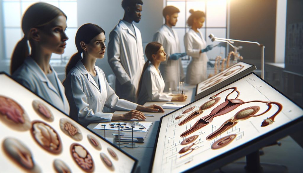
Discover the combining form for reproductive glands and unlock the secrets to understanding fertility. Get the insights you need for your journey today!

Discover why cats have barbed reproductive organs and what it means for feline anatomy. Uncover the secrets of your pet’s biology and enhance your understanding today!

Discover how the women’s reproductive system works in this essential guide. Learn about fertility, treatment options, and emotional support on your journey.

Discover how the reproductive system maintains homeostasis effectively! Learn its crucial role in fertility and health-uncover the secrets to balancing hormones today!

Discover essential tips on how to take care of your reproductive system to boost fertility and overall health. Don’t miss these vital insights for a healthier future!

Unlock the mysteries of the female reproductive system! Discover its key parts, functions, and how they affect fertility. Learn more to empower your health journey.
Discover how a septate hymen can lead to infertility. Learn about this hidden barrier to conception and find out what steps you can take today for a hopeful future.